
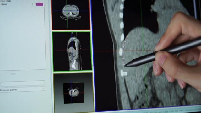
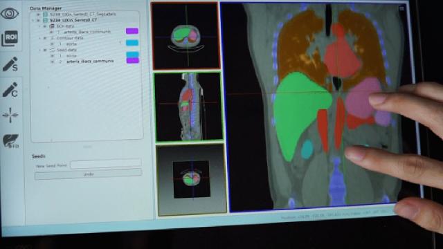
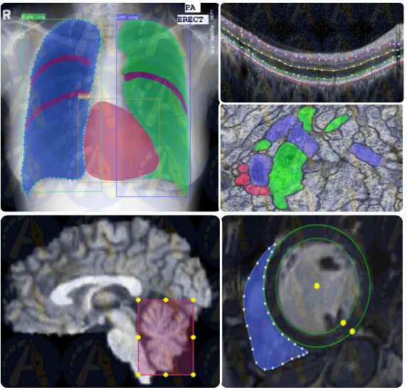
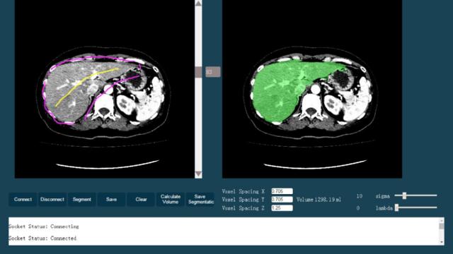
AS (Anatomy Sketch) software
is an open source software for medical image analysis developed by
the Medical Imaging Research Group of Dalian University of Technology
, aiming at scientific research colleagues and providing convenience tools. In the process of medical image research, it is often necessary to browse, process and mark the image. AS wants to act as a kind of clean and extensible software platform meeting this need, supporting basic image and mesh processing and quick annotation using mouse or stylus, and allowing users to plug in custom Python or DLL function module (including deep neural network). Software (
Download, unzip, double-click AnatomySketch.exe
), constant updates in GitHub (
webpage
and
download
), the software's main menu has the "Help" button with detailed software description and demo videos. One of the two neural network examples with pythoy plugin is Abdomonial CT (G
itHub webpage
,
local download
). Please send email to
mingruizhuang@foxmail.com
.
;)
;)
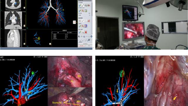
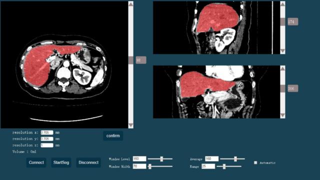
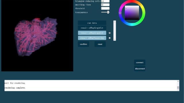
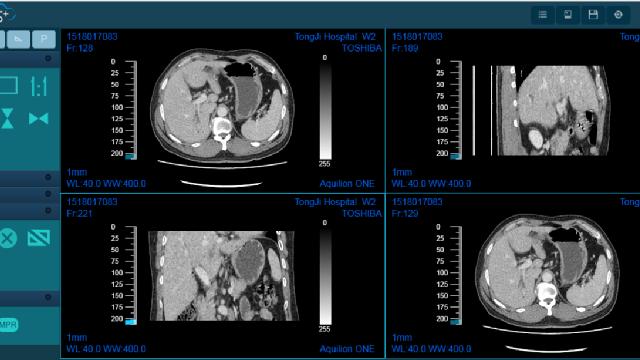
DeepInsight system
is a pulmonary precision surgery assistance system jointly developed by
the Key Laboratory of Medical Imaging Intelligent Computing of The Ministry of Education (Northeast University)
and the thoracic Surgery team of Jiangsu Provincial People's Hospital, providing functions such as three-dimensional bronchus and vascular reconstruction, precise nodule positioning and preoperative planning. At present the system is free of charge to the general thoracic surgeons. For download and instructions, please refer to the "DeepInsight system" WeChat (
attention into the official account
).
Trial application instructions
are included in the "instructions", data download (
software installation file, the application protocol, installation instructions, usage instructions, etc.
), In the WeChat official account, there is chest 3D reconstruction image acquisition quality advice (
download
). Please send email to
tanwenjun@cse.neu.edu.cn
.
;)
;)
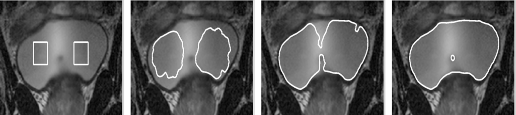
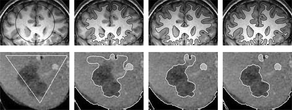
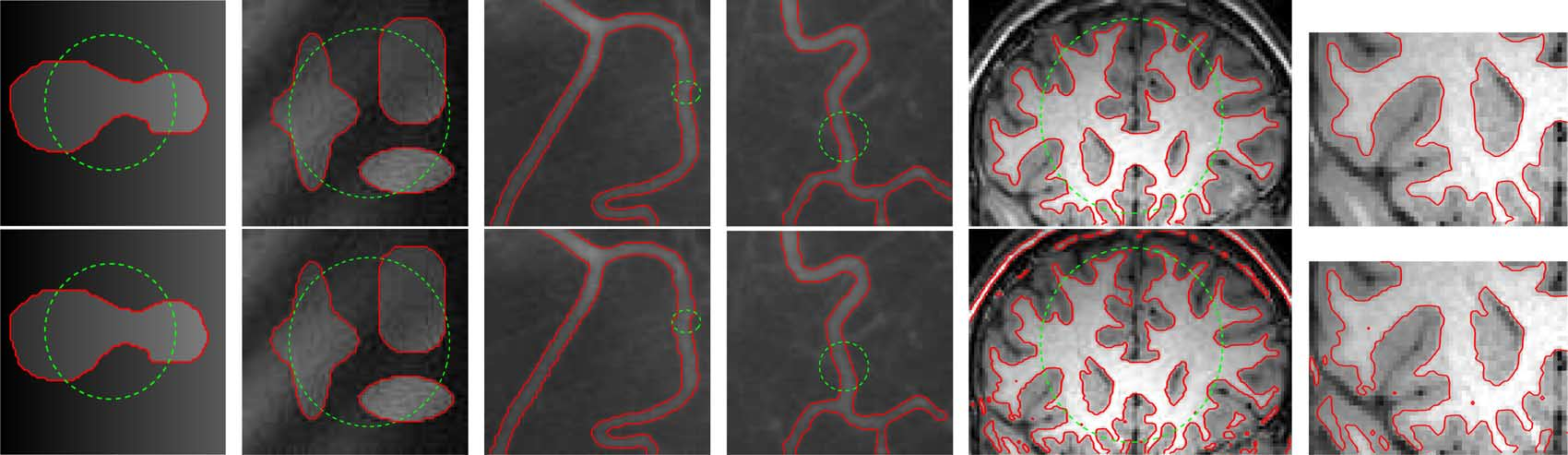
DRLSE algorithm package
is a image segmentation software developed by
Professor Li Chunming's team of University of Electronic Science and Technology of China
, DRLSE algorithm was defined as Distance Regularized Level Set Evolution and applied to
an edge-based active contour model for image segmentation
with fast computing efficiency. The famous paper was published in 2010 by Dr. Li and has been cited more than 1200 times. DRLSE won IEEE Signal Processing Society Best Paper Award in 2015 (
paper download
). The algorithm code was written in Matlab and open for download (
code download
), Please send email to
chunming.li@uestc.edu.cn
.
;)
;)
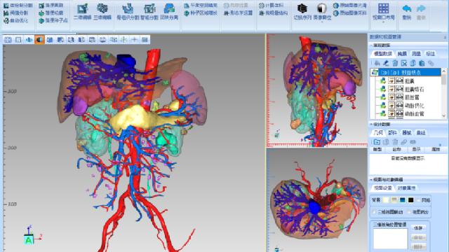
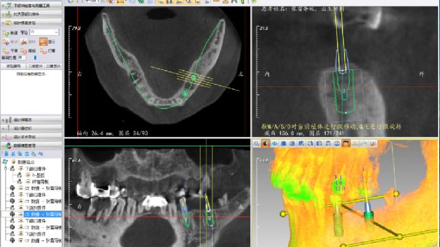
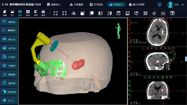
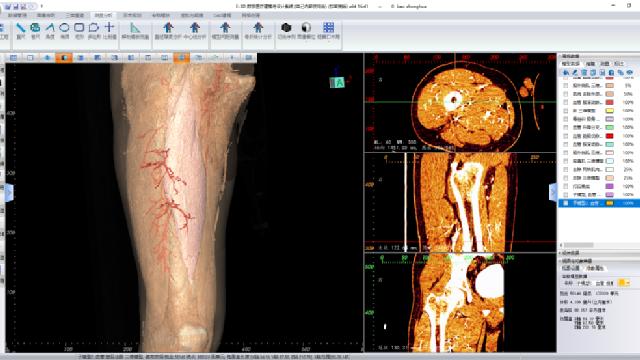
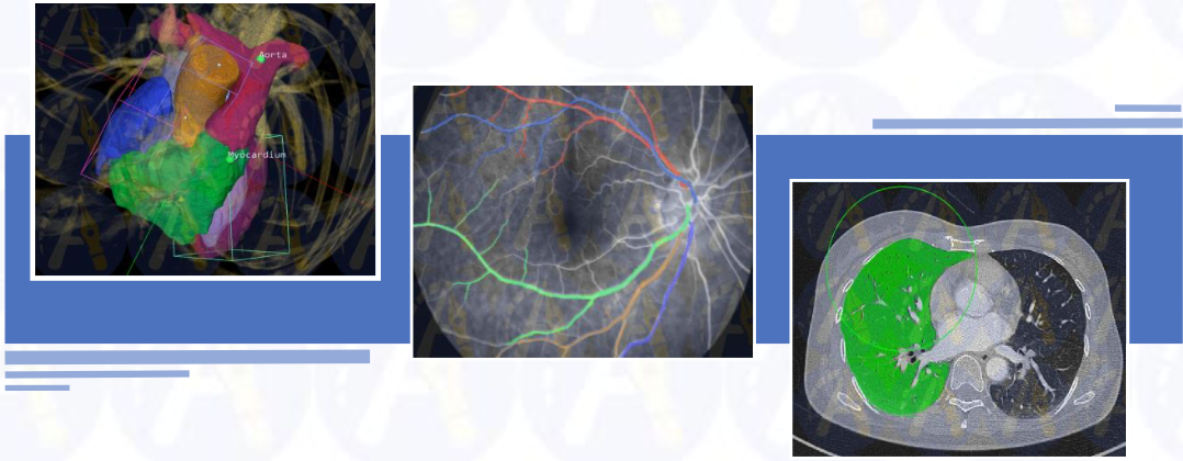
E3D 3D Digital Medical Modeling and Design system
was developed by
Professor Liao Shenghui's team from the National Engineering Laboratory of Medical Big Data Application Technology
, Central South University. The sytem supports intelligent image segmentation and 3D reconstruction, mesh processing, surgical planning, model automatic registration, mirror repair, nail path design, fracture reduction, 3D vascular analysis, fracture line cloud map analysis, CAD 3D modeling, surgical guide plate design, measurement and annotation. Intelligent image segmentation can segment 10 kinds of tissue targets at one time. Support multi-mode image automatic registration and fusion; Support CT-based simulation of X-ray positive and negative images; Support real-time dynamic 3d osteotomy effect; Support automatic repair of grid model with one key. The surgical guide plate supports the modular design and multiple reuse of the nailing guide, grooving guide, bridge structure and motherboard. The system supports the operations of the guide plate such as hollow out, adding text and cutting edge. CAD sketch design can be carried out, and CAD models can be generated using rotation, drawing and other operations. It provides remote cloud platform interface for 3D display and communication on mobile phones. There are abundant learning resources (
system download, operation manual, video tutorial
) on the software website. Please send email to
lsh@csu.edu.cn
.
;)
;)
;)
;)
;)
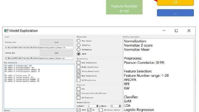
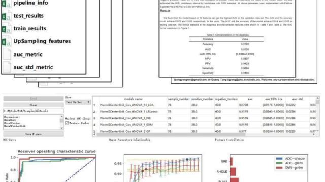
FAE (FeAture Explorer) software
is an open-source software developed by
Shanghai Key Laboratory of Magnetic Resonance, East China Normal University
. In projects of
Radiomics
, researchers need to extract quantitative features from medical images and to build machine-learning models. Although there are several software helping build models, researchers have to spend much time on fine-tuning different algorithms and hyper-parameters. FAE is being developed to optimize the process in Radiomics project. FAE includes four major parts, i.e. Feature Extraction, Data Preprocessing, Model Development, and Result Description. FAE provides a user-friendly UI to help researchers, especially who are not familiar with machine learning or coding, complete a project of Radiomics. Furthermore, FAE can develop thousands of models with one-click, compare the results from these models, and find the optimal model for the specific project. All results generated during the model development are stored and allows researchers to use them in other software. The source code of FAE is on
Github
. The release version of Windows 64bit can be directly downloaded locally (
download, unzip and double-click FAEv0.3.6\MainFrameCall\MainFrameCall.exe file
), which including the demo,
manual document
, and
research paper published in PlusOne
. Online videos can teach you how to easily use FAE software (
video link
). Please send email to
songyangmri@gmail.com
.
;)
;)
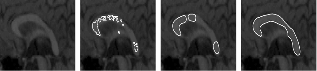
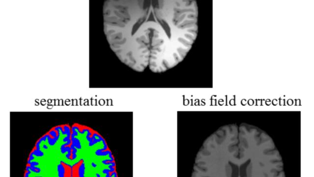
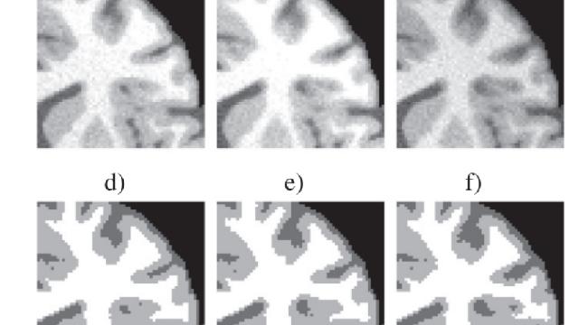
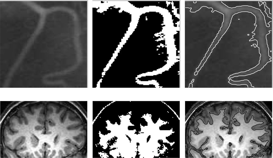
MICO software package
is a medical image segmentation software developed by
Professor Li Chunming's team of University of Electronic Science and Technology of China
, which is used for brain tissue segmentation and partial field estimation or correction of normal MR image data. MICO is the abbreviation of Multiplicative intrinsic component optimization. It is an open source algorithm based on Matlab published by Professor Li Chunming in 2014. The MICO algorithm has been validated on many MRI images and shows advantages over many popular algorithms in terms of robustness, efficiency, and accuracy. Matlab code is clear and concise, easy to read and understand. Users and developers can easily work on applications for further development.
MICO software package (
compressed file download
,
Toolkit download
,
MathWorks website introduction
). Paper was published in 2014 (
paper download
), Please send email to
chunming.li@uestc.edu.cn
.
;)
;)
Pair
is the first
one-stop medical image annotation software
in China. It is compatible with all image modes and formats, supports multiple annotation types, and provides A auxiliary annotation tool. Users can highly customize file configuration and workflow. Up to now, Pair has been used by more than 30 hospitals and 500 users, and has been highly praised for undertaking more than 400 target-marking tasks.
Pair packages (
Compressed file download
), contact email address:
pair_all_purp_anno@163.com
.
;)
;)
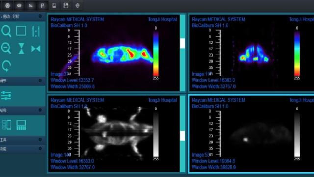
RayPlus software
is a medical image recognition and processing software developed by the
Digital PET Laboratory of Huazhong University of Science and Technology
. RayPlus is a cloud-end intelligent medical imaging platform. Combined with intelligent medical image processing technology and cloud computing technology, RayPlus provides doctors with lightweight cloud CAD&T service to assist medical decision-making for major diseases and to improve the treatment effect of patients with severe diseases. The next step will be to include data from PET pulse Raw Data as AI input, instead of being restricted to the final medical image, the system website is
http://www.rayplus.top
, the software instructions (
local download
). Please send email to
petlab@mail.hust.edu.cn
.
;)
;)
;)
;)
;)
RSF algorithm package
is a image segmentation software developed by
Professor Li Chunming's team of University of Electronic Science and Technology of China
, RSF energy was defined as Region-Scalable Fitting Energy and minimized for image segmentation. The famous paper was published in 2008 by Dr. Li and has been cited 1876 times. RSF won IEEE Signal Processing Society Best Paper Award in 2013 (
paper download
). The algorithm code was written in Matlab and open for download (
code download
), Please send email to
chunming.li@uestc.edu.cn
.
;)
;)
;)
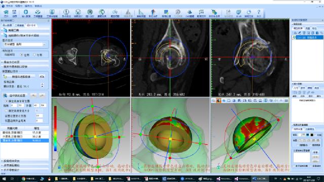
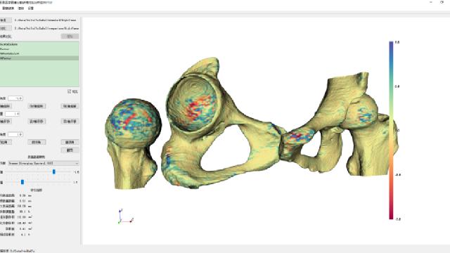
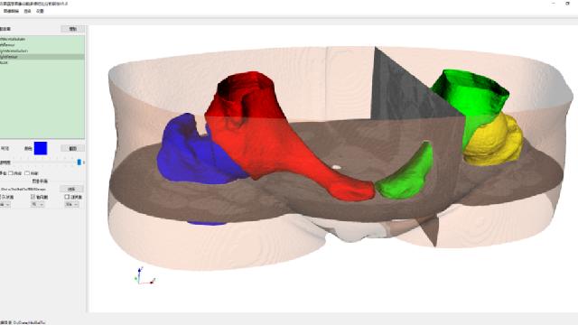
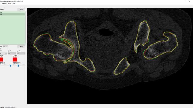
VolumeShop_Compare software
is a medical imaging AI software developed by Nanjing HuiBaiTu Technology Co., LTD. and is part of the intelligent image segmentation and comparison analysis function module of VolumeShop medical imaging AI software (under development). VolumeShop_Compare realizes 2D and 3D visual comparative analysis of medical image segmentation. VolumeShop_Compare assists researchers in experimental analysis of medical image segmentation made by AI algorithms. VolumeShop_Compare mainly the following functions: (1) 3D visualization of surface distance difference between the segmentation result of the algorithm and the gold standard of the doctor's label, (2) visually comparison of the difference between the segmentation result of the algorithm and the gold standard of the doctor's label on multi-orientation slices, (3) 3D visualization rendering of the segmentation result. There are Windows version software (
local download of VolumeShop_Compare 1.0.0.msi
), software instructions (
operation manual PDF download
), and demo data including pelvic CT images, standard masks and AI segmented masks (
Testdata zip download
). Please send email to
changyong@huibaitu.com
。
;)
;)
;)
www.cvnis.net, All Rights Reserved.
本站所有内容版权所有,未经许可请勿转载。
本站所有内容版权所有,未经许可请勿转载。